I. Core Factors Influencing Clinical Laser Efficacy – Operator Self-Check Checklist
Identical equipment and identical protocols — why are clinical outcomes so variable? Please review against the checklist below:
For Soft Tissue Cutting
| No. | Check Item | Yes / No |
|---|---|---|
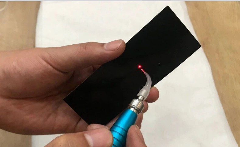
| 1 | Is the fiber tip carbonized? | ☐ |
| 2 | Are you using puncture incision technique (rather than dragging incision)? | ☐ |
| 3 | Have you marked the position with a probe in advance? | ☐ |
| 4 | Is the cutting time kept within a reasonable range? (Longer duration leads to greater thermal injury) | ☐ |
For Hemostasis (Bleeding Control)
| No. | Check Item | Yes / No |
|---|---|---|
| 1 | Is the fiber tip carbonized? | ☐ |
| 2 | Have you located the precise bleeding point (instead of merely coagulating surface blood clots)? | ☐ |
| 3 | Is the fiber tip in direct contact with tissue/blood vessels (not hovering above the blood)? | ☐ |
| 4 | Is the bleeding inflammatory? (If yes, remove granulation tissue first) | ☐ |
For Physical Therapy (Low-Level Laser Therapy)
| No. | Check Item | Yes / No |
|---|---|---|
| 1 | Is the fiber tip non-carbonized (kept clean)? | ☐ |
| 2 | Is the treatment performed in non-contact mode (approximately 1 cm away from tissue)? | ☐ |
| 3 | Does the patient experience mild warmth and comfort (no burning or pain)? | ☐ |
| 4 | Is the fiber tip kept in continuous motion (not stationary at one spot)? | ☐ |
| 5 | Has local anesthesia been avoided (patient feedback is required)? | ☐ |
Periodontal Pocket Treatment
| No. | Check Item | Yes / No |
|---|---|---|
| 1 | Has the causative factor (dental calculus) been eliminated? | ☐ |
| 2 | Is the correct mode selected — disinfection (non-carbonized) or debridement (carbonized)? | ☐ |
| 3 | Are fiber movements predominantly horizontal (left-right sweeping)? | ☐ |
| 4 | For hemostasis, is treatment only applied at the opening of the periodontal pocket (not deep within the pocket)? | ☐ |
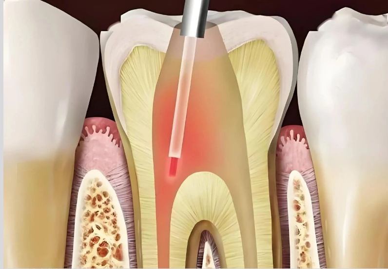
For Root Canal Disinfection
| No. | Check Item | Yes / No |
|---|---|---|
| 1 | Are you using a 200 μm optical fiber (thin enough to access the root canal)? | ☐ |
| 2 | Is the fiber tip non-carbonized? | ☐ |
| 3 | Is the laser procedure combined with conventional root canal preparation and irrigation? | ☐ |
Second-Stage Implant Uncovering
| No. | Check Item | Yes / No |
|---|---|---|
| 1 | Have you precisely located the implant with a probe in advance? | ☐ |
| 2 | Are you using intermittent footswitch activation + pulsed mode + saline cooling? | ☐ |
| 3 | Is the gingival thickness appropriate? (Excessively thick or thin gingiva will lead to complications) | ☐ |
For Dentin Desensitization
| No. | Check Item | Yes / No |
|---|---|---|
| 1 | Is the indication exposed dentin (not pulpitis)? | ☐ |
| 2 | Have you marked with a 2B pencil and maintained a dry tooth surface? | ☐ |
| 3 | Has no local anesthesia been administered? (Patient feedback is essential) | ☐ |
| 4 | Have you informed the patient that desensitization treatment is not permanent? | ☐ |
II. Common Errors and Corrective Actions
| Error | Correction |
|---|---|
| Error 1: Either never carbonizing the fiber, or carbonizing for every procedure | Cutting, debridement, coagulation → carbonize. Disinfection, physiotherapy, root canal disinfection → do NOT carbonize. |
| Error 2: Dragging the fiber while cutting soft tissue, taking too long to sever | Use perforation-link cutting – dense perforations then connect them. Shorter time, less thermal damage. |
| Error 3: Coagulating only the surface clot | Press with a cotton pellet → locate the bleeding point → directly contact the vessel/tissue with the carbonized tip → hold for 2–3 seconds. |
| Error 4: Setting the power too high for physiotherapy | Physiotherapy requires low energy to stimulate cell growth. Too high, and it turns from “stimulation” into “destruction.” |
| Error 5: Using laser alone for periodontal treatment without scaling | Laser cannot remove calculus. Without removing the etiology, recurrence is inevitable. Laser is adjunctive, not replacement. |
| Error 6: Using laser to coagulate the extraction socket after tooth extraction | Bone tissue is heat-sensitive – laser coagulation damages bone. Post-extraction laser is for soft-tissue physiotherapy only. |
| Error 7: Treating lichen planus or leukoplakia with laser | These are potentially malignant conditions. At the general practice level, do not treat them – the risk-benefit ratio is unacceptable. |
| Error 8: Administering local anesthesia for desensitization | Desensitization relies on patient feedback (“does it hurt?”) to guide the endpoint. No feedback → risk of over-irradiation. |
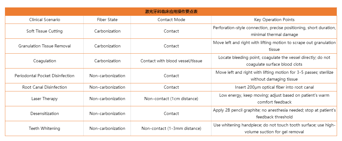
III. One-Sentence Summary for Each Clinical Scenario
IV. Clinical Safety Precautions
1. Eye Protection
| Requirement | Action |
|---|---|
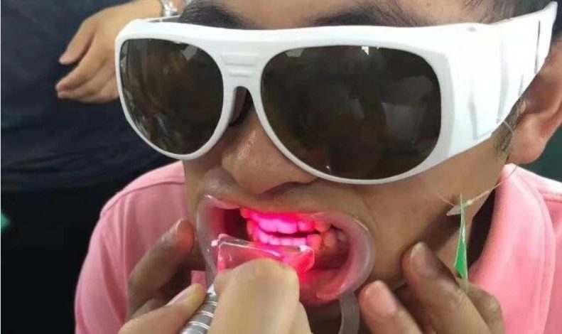
| All personnel (operator and patient) must wear protective eyewear | Do not skip this step |
| Laser warning sign must be posted in the treatment area | Visible to everyone entering |
| Never direct the laser toward the eyes | Even with closed eyelids – the beam can still cause damage |
2. Thermal Damage Prevention
| Principle | Implementation |
|---|---|
| Do not stay in one spot for too long | Keep the fiber moving continuously |
| Be extra cautious with heat-sensitive structures | Implants, bone tissue, and thin gingiva |
| Use saline irrigation when necessary | Particularly suitable for cases of implant exposure and prolonged surgical procedures. |
3. Reflected Beam Protection
| Scenario | Note |
|---|---|
| Intraoral metal crowns, exposed implants or other reflective surfaces | The intensity of reflected beams increases significantly |
| All personnel present | Protective goggles must also be worn. |
4. Whitening Gel Protection
| Risk | Prevention |
|---|---|
| Whitening gel contacting the mucosa | This can cause burns and ulcers |
| Gingival protection | The gingival barrier must be applied thoroughly and light-cured |
| Gel removal | Use high-volume suction. Never wipe with cotton balls or gauze, as this will spread the gel. |
5. Post-treatment Follow-up
| Action | Purpose |
|---|---|
| Document each laser treatment | Track outcomes and adjust protocols |
| Conduct follow-ups for recurrent conditions (periodontitis, gingival tumors, etc.) | Long-term management is critical |
| Set realistic patient expectations | Do not promise a “one-time cure” — remai |
Semiconductor lasers have been used in dentistry for many years. They have evolved from an initial “novelty” into a routine clinical tool. Its greatest advantage lies in the fact that once practitioners master the basic principles and proper operating techniques, a single device can meet treatment requirements across multiple dental specialties.
Nevertheless, its effectiveness always relies on proper understanding and correct operation. With identical equipment and identical power settings, operational variables including carbonized versus non-carbonized fiber tips, contact versus non-contact modes, and high versus low energy settings can lead to drastically different outcomes.
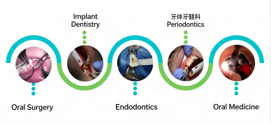
We hope this three-part series — ranging from fundamental principles to clinical procedures — can help fellow practitioners avoid common pitfalls in daily practice and fully unlock the potential of the lasers they use.